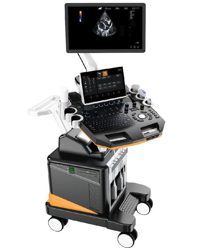
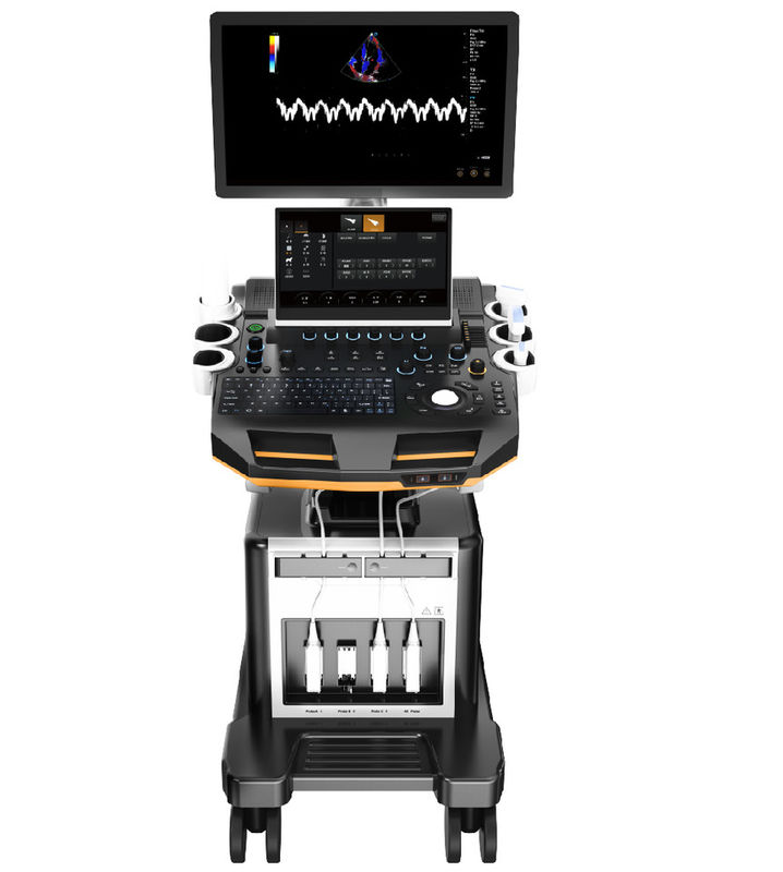
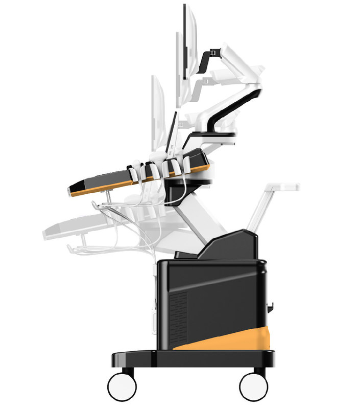
| 1. Veterinary Color Doppler Ultrasound System |
| 1.1 Main structure:Trolley type, double screen |
|
2. Applications and Report
2.1 To meet the examination and diagnosis needs of pet hospital and scientific research institutions in the digestive system, reproductive system, urinary system, physical examination, etc.
|
|
3. Main Specifications
3.1. Full Digital Color Doppler Ultrasonic Mainframe
|
| 3.2. Digital Beam Enhancer |
| 3.3. Multiple-beam synthesis |
| 3.4. Full-Digital 2D gray scale imaging |
| 3.5.Tissue harmonic imaging(THI) |
| 3.6.B/C Real-time two synchronous imaging |
| 3.7.M mode imaging |
| 3.8.Anatomic M mode imaging (Sampling line≥3) |
| 3.9.Color Doppler imaging(C,PDI,DPDI) |
| 3.10.Spectral Doppler imaging(PW,HPRF PW,CW) |
| 3.11.Tissue Doppler imaging (TVI, M-mode, spectral imaging, etc.) |
| 3.12.Four-dimensional supersonic image formation |
| 3.13.★Contrast Tuned Imaging (optional) |
| 3.14.★PView wide field imaging (optional) |
| 3.15.Space compound imaging (application to Abdominal, GYN, vessel, superficial small organs, can be dual image contrast display at same time) |
| 3.16.Frequency & Focus Compound |
| 3.17.Extended field-of-view(EFOV) |
| 3.18.Real-time dual image contrast display |
| 3.19.B/C/D Real-time three synchronous imaging |
| 3.20.Speckle Reduce imaging(SRI) |
| 3.21.Elastography(optional) |
| 4.★Operation Interface: support 10 different languages |
|
5. System Specification
5.1 Monitor:21.5 inch high resolution medical LCD monitor, adjustable angle
13.3 inch touch screen, support multi-touch
|
| 5.2 ★Probe connectors: ≥4 active, support all probes |
|
5.3.2D imaging mode
1)Full Digital Beamformers
2)Digital full dynamic focusing, digital variable aperture and dynamic trace change, A/D≥15 bit
3)Receiving mode: transmitting and receiving channel ≥1024, multi-signal parallel processing
4)Line density: ≥512, visible and adjustable
5)Under B mode, focus number≥10 level, focus position continuously adjustable
6)TGC:8 segment adjustable
7)Gain: B/M/D separately adjustable,≥100dB
8)★Dynamic range:≥180dB
9)★Max scanning depth:≥360mm
10)Grey scale:0-67, visible and adjustable
11)Sound power:1%-100%
12)Linear array probe two-dimensional independent deflection
13)ZoomPIP(1.5 / 2.0 / 2.5 / 3.0 / 3.5 / 4.0 / 4.5 / 5.0 / 10 times)
|
|
5.4. Color Doppler Imaging mode
1)Imaging: including velocity, velocity variance, energy, direction energy display, etc
2)Display mode:B/C,B/C/M,B/POWER,B/C/PW
3)Line density≥3 level
4)Color hiding technology: without back to B mode can hide C mode, only color speed ruler display
5)Blood flow graph,color flow profile to measure intravascular velocity
|
|
5.5. Spectral Doppler Imaging
1)Display: full screen, duplex / triplex (PW only)
2)Gain:≥100dB
3)Velocity ≥4 level adjustable
4)Max scanning speed:PWD:Positive or negative blood flow velocity≥7.6m/s;
CWD:blood flow velocity≥20.0m/s,min:≤5 mm /s(Nonnoise signal);
5)Zero shift adjustable ≥8 level
6)Display mode:B, PW, B/PW, B/C/PW, B/CW, B/C/CW, etc
7)Spectrum envelope function, Manual Measurement
8)Display control: reverse, zero shift, B refresh, D extension, B/D extension, etc
9)Smart Doppler technology: freely switch between real-time B+CFM mode and real-time PW mode
|
|
5.6.★PView wide field imaging (optional)
1)High-resolution display, length can up to 50cm
2)Imaging support forward erasure, no need to re-imaging
3)With two dimensional wide view imaging mode and color wide view imaging mode
|
|
5.7.Probe: wideband with frequency conversion, independent frequency conversion under B and CFM
1)Fundamental Frequency conversion≥3 level;
2)1.Convex probe
Fundamental frequency:nine levels frequency conversion
2.0MHz/2.3MHz/2.5MHz/3.0MHz/3.5MHz/4.0MHz/4.6MHz/5.0MHz/5.4MHz,
Harmonic Frequency:4.0MHz/4.6MHz/5.0MHz,three levels frequency conversion
2. Linear probe
Fundamental frequency:nine levels frequency conversion 4.0MHz/4.6MHz/5.0MHz/6.0MHz/7.0MHz/8.0MHz/9.2MHz/10.0MHz/12.0MHz/13.3MHz
Harmonic Frequency:8.0MHz/9.2MHz/10.0MHz,three levels frequency conversion
3. Phased array probe
Fundamental frequency:nine levels frequency conversion 1.7MHz/1.9MHz/2.1MHz/2.5MHz/3.0MHz/3.4MHz/3.8MHz/4.2MHz/5.0MHz
Harmonic Frequency:3.4MHz/3.8MHz/4.2MHz,three levels frequency conversion
4. High frequency phased array probe
Fundamental frequency: eight levels frequency conversion: 3.0MHz/3.5MHz/4MHz/5MHz/5.4MHz/6MHz/7MHz/8MHz,
Harmonic Frequency: 6MHz/7MHz/8MHz,three levels frequency conversion
5.8 Micro-convex probe
Fundamental frequency:eight levels frequency conversion
3.0MHz/3.5MHz/4.0MHz/5.0MHz/5.4MHz/6.0MHz/7.0MHz/8.0MHz
Harmonic Frequency: 6.0MHz/7.0MHz/8.0MHz,three levels frequency conversion
|
|
5.9 Technical requirements for electronic convex probe:
Frequency:2.0-5.4 MHz,lateral resolution: ≤2 (depth≤80), ≤3mm (80<depth≤130)
axial resolution: ≤1 (depth≤80), ≤1mm (80<depth≤130)
dead zone:≤3mm; Geometric position accuracy: broadwise:≤5%; lengthwise:≤5%
detecting depth≧240 mm
|

 Your message must be between 20-3,000 characters!