logo
Home > Products > Veterinary Ultrasound Machine >
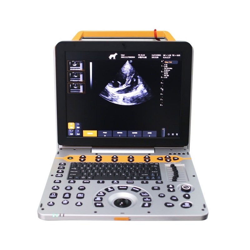
15 inch P60V Portable Veterinary Color Doppler Ultrasound System for Small Animal Clinics

15 inch P60V Portable Veterinary Color Doppler Ultrasound System for Small Animal Clinics

15 inch veterinary color Doppler ultrasound

portable ultrasound system for small animals

P60V veterinary ultrasound machine with warranty

Place of Origin:

China

Brand Name:

Golead

Certification:

ISO13485

Model Number:

P60V

Contact Us

Request A Quote
Product Details
Product Name:
15 Inch Portable Veterinary Ultrasound Machine For Small Animals
Display:
15'' LCD
Element:
128
Channel:
64
Display Mode:
B, PW, B/PW, B/C/PW, B/CW, B/C/CW
Battery:
13,600mAh
Continuous Scanning Time:
> 1.5 Hours
Gray Scale:
256
Payment & Shipping Terms
Minimum Order Quantity
1 Unit
Packaging Details
carton box
Delivery Time
2 working days
Payment Terms
T/T
Supply Ability
1,000 units per month
Product Description

15 inch P60V Portable Veterinary Color Doppler Ultrasound System for Small Animal Clinics
 
Overview
Portable veterinary ultrasound machine P60V used advanced and stable PC system, combing excellent images, all-round functions, rich image management software together to meet the needs of various animal groups in different environments. The built-in battery with large capacity imported chip, dual probe interface, canbe configured with a variety of probes to satisfy the needs of different examination modes
 

The P60V Portable Veterinary Color Doppler Ultrasound System is designed for comprehensive diagnostic imaging in veterinary hospitals, specialty clinics and teaching institutions.

Equipped with advanced beamforming technology, Tissue Harmonic Imaging (THI), Color Doppler and multiple probe options, the system supports abdominal, cardiac, reproductive, musculoskeletal and vascular examinations in companion animals.

Its portable design, dual active probe connectors and long battery life allow veterinarians to perform high-quality examinations both in clinics and during mobile visits.


Features

Multiple Beam Synthesis

Improves image uniformity and spatial resolution, helping veterinarians visualize anatomical structures more clearly.


Tissue Harmonic Imaging (THI)

Enhances contrast resolution while reducing image noise, supporting clearer visualization of abdominal organs.


Speckle Reduction Imaging (SRI)

Helps reduce speckle artifacts and improve tissue border definition.


Dual Active Probe Connectors

Allows fast switching between probes without repeated reconnection, improving workflow efficiency.


15-inch Medical LCD

Provides a larger viewing area for detailed image review during routine examinations.


Parameters

1. Product Name: 15 inch Portable Vet Ultrasound Machine for Small Animals 
1.1. Structure Style: Portable

2. Application
2.1. Applicable for animals’ examination and diagnosis on digestive system, reproductive system, urinary system for animal hospitals and scientific institutions.

3. System Technical Specifications and Summary
3.1. Full digital color doppler ultrasound diagnostic system

3.2. Digital Beam Enhancer
3.3. Multiple Beam Synthesis
3.4. Full-Digital 2D Gray Scale Imaging
3.5. Tissue Harmonic Imaging (THI)
3.6. B/C Real-time Two Synchronous Imaging
3.7. M Mode Imaging
3.8. Anatomic M Mode Imaging (Sampling line≥3)
3.9. Color Doppler Imaging (CFM, PDI, DPDI)
3.10. Spectral Doppler Imaging (PW, HPRF PW, CW)
3.11. Tissue Doppler Imaging (TVI, M-mode, Spectral Imaging, etc.)
3.12. Four-dimensional Supersonic Image Formation
3.13.★Contrast Tuned Imaging (optional)
3.14.Wide Field Imaging (optional)
3.15. Space compound imaging (application to Abdominal, GYN, vessel, superficial small organs, can be dual image contrast display at same time)
3.16. Frequency & Focus Compound
3.17. Extended field-of-view (EFOV)
3.18. Real-time dual image contrast display
3.19. B/C/D Real-time three synchronous imaging
3.20. Speckle Reduce Imaging (SRI)
3.21. Elastography (optional)
4.★Operation Interface: support 10 different languages

5. System Specification
5.1. Monitor: 15 inch high resolution medical LCD monitor

  1. ★Probe connectors: ≥2 active, support all probes

5.3. 2D Imaging Mode
1)Full Digital Beamformers
2)Digital full dynamic focusing, digital variable aperture and dynamic trace change, A/D≥15 bit
3)Receiving mode: transmitting and receiving channel ≥1024, multi-signal parallel processing
4)Line density: ≥512, visible and adjustable
5)Under B mode, focus number≥10 level, focus position continuously adjustable
6)TGC: 8 segment adjustable
7) Gain, ≥100dB
8)★Dynamic range ≥180dB
9)★Max scanning depth ≥360mm
10)Grey scale: 0-67, visible and adjustable
11)Sound power: 1%-100%
12)Linear array probe two-dimensional independent deflection
13)Zoom(1.5 / 2.0 / 2.5 / 3.0 / 3.5 / 4.0 / 4.5 / 5.0 / 10 times)

5.4. Color Doppler Imaging Mode
1)Imaging: including velocity, velocity variance, energy, direction energy display, etc
2)Mode: B/C, B/C/M, B/POWER, B/C/PW
3)Line density ≥3 level
4)Color hiding technology: without back to B mode can hide C mode, only color speed ruler display
5)Blood flow graph, color flow profile to measure intravascular velocity

5.5. Spectral Doppler Imaging
1)Display: full screen, duplex / triplex (PW only)
2)Gain ≥100dB
3)Velocity ≥4 level adjustable
4)Max scanning speed: PWD: Positive or negative blood flow velocity≥7.6m/s;
CWD: blood flow velocity ≥20.0m/s, min ≤5 mm/s (Nonnoise signal);
5)Zero shift adjustable≥8 level
6)Display mode: B, PW, B/PW, B/C/PW, B/CW, B/C/CW, etc
7)Automatic Measurement, Manual Measurement
8)Display control: reverse, zero shift, B refresh, D extension, B/D extension, etc
9)Smart Doppler technology: freely switch between real-time B+CFM mode and real-time PW mode

5.6. ★PView wide field imaging (optional)
1)High-resolution display, length can up to 50cm
2)Imaging support forward erasure, no need to re-imaging
3)With two dimensional wide view imaging mode and color wide view imaging mode

5.7. Probe: wideband with frequency conversion, independent frequency conversion under B and CFM
1)Fundamental Frequency Conversion≥3 level;
2)1. Convex probe
Fundamental frequency: nine levels frequency conversion
2.0MHz/2.3MHz/2.5MHz/3.0MHz/3.5MHz/4.0MHz/4.6MHz/5.0MHz/5.4MHz,
Harmonic Frequency: 4.0MHz/4.6MHz/5.0MHz, three levels frequency conversion
2. Linear probe
Fundamental frequency: nine levels frequency conversion 4.0MHz/4.6MHz/5.0MHz/6.0MHz/7.0MHz/8.0MHz/9.2MHz/10.0MHz/12.0MHz/13.3MHz
Harmonic Frequency: 8.0MHz/9.2MHz/10.0MHz, three levels frequency conversion
3. Phased array probe
Fundamental frequency: nine levels frequency conversion 1.7MHz/1.9MHz/2.1MHz/2.5MHz/3.0MHz/3.4MHz/3.8MHz/4.2MHz/5.0MHz
Harmonic Frequency: 3.4MHz/3.8MHz/4.2MHz,three levels frequency conversion
4. High frequency phased array probe
Fundamental frequency: eight levels frequency conversion:
3.0MHz/3.5MHz/4MHz/5MHz/5.4MHz/6MHz/7MHz/8MHz,
Harmonic Frequency: 6MHz/7MHz/8MHz,three levels frequency conversion
5. Micro-convex probe
Fundamental frequency: eight levels frequency conversion
3.0MHz/3.5MHz/4.0MHz/5.0MHz/5.4MHz/6.0MHz/7.0MHz/8.0MHz
Harmonic Frequency: 6.0MHz/7.0MHz/8.0MHz, three levels frequency conversion

5.8. Technical requirements for electronic convex probe:
Frequency: 2.0-5.4 MHz, lateral resolution: ≤2 (depth≤80), ≤3mm (80<depth≤130)
axial resolution: ≤1 (depth≤80), ≤1mm (80<depth≤130)
dead zone: ≤3mm; Geometric position accuracy: broadwise: ≤5%; lengthwise:≤5%
detecting depth≧240 mm

5.9. Technical requirements for electronic linear probe:
Frequency: 4.0-13.3MHz, lateral resolution: ≤1 (depth≤40),
axial resolution: ≤0.5 (depth≤50),
dead zone: ≤3mm; Geometric position accuracy: broadwise:≤4%; lengthwise:≤2%
detecting depth≧100 mm

6. Measurement / Analysis and Reports
6.1. General measurement: distance, area, ellipse, cross line, angle, distance ratio, volume, volume (ellipse), area ratio, diameter, joint angle.

6.2. Specialized measurement:
1) Cardiac: Automatic spectrum envelope, LV, Main Pulmonary artery diameter, RVEDd, RVEDs, LVM, LAV, HR, MVF, AO, AR, LVOT, TVF, Pulmonic valve, Pulmonary vein, RV, Doppler fetal heart sound, LVET, LVM, LVMI, AV
2) Vascular: carotid intima (IMT), length stenosis ratio, area stenosis ratio, IMT (back wall), IMT (front wall)
3)★OB: Canine: GSD, CRL, HD, BD; Feline: BD, HD; Swine: HLA, SLA;
Bovine: CRL, BTD, BUD; Ovine: SCRL, BPD; Equine: GSD(H), ESD (V)
4) Urinary measurement: Prostate, residual urine, left kidney, right kidney, left adrenal gland,
right adrenal gland, left testis, right testis, left seminal vesicle, right seminal vesicle.

  1. Abdomen measurement: liver, common hepatic duct, diameter of portal vein, gallbladder, common

bile duct, pancreas, spleen, abdomen aortic diameter, kidney.
6)Organella.

7. Peripherals Section
7.1. Configured ultrasonic graphic work station, the work station software equipped with registration certificate, supports black and white digits, simulation B/W, digit color, simulation color, text and video printer, and foot switch
7.2. Support Internet connection
7.3. Support DICOM 3.0, report of DICOM Obstetrics and Gynecology / Heart / Blood Vessel
7.4. Video/Audio Input Output
7.5. Main unit comes with USB port
7.6. Support ultrasonic system directly sent clinical images and reports through internet

8. Cine Loop and Original Data Processing
8.1. Cine Loop ≥ 3061 frames, support manual and auto operation
8.2. Original Data Processing, can conduct offline parameters analysis on static files and replay dynamic images. Such as Gain, Pseudo Color, Grey Scale Curve, ect.
8.3. Digitalization Hard Drive Capacity ≥ 256G, support permanent store static, dynamic images, can be consulted, transferred, deleted freely
8.4. Multiple Image Export Modes: Dynamic Image and Static Image exported in PC format, Images can be seen on regular PC devices, no need of specific software.
can process diagnosis, export and backup image data at the same time without affecting the inspection operation
8.5. Professional probe holder ≥2 (coupling holder not included)

9. After-Sale Service
9.1. Mani unit warranty ≥2 years
9.2. Lifelong maintenance after warranty, lifelong free updates service of software
9.3. Free regular return visit for maintenance
9.4. If any repair reported, we will reply and propose solution within 24 hours.


Quality Assurance

Each ultrasound system undergoes factory testing before shipment, including:

  • Image quality verification
  • Probe performance testing
  • Doppler functionality testing
  • Electrical safety inspection
  • Display calibration
  • Battery performance verification
  • Software functional testing
  • Packaging inspection

Every unit is tested before shipment to help ensure reliable clinical performance.


Advantages

Why Choose P60V?

  • High-resolution color Doppler imaging
  • Dual active probe connectors
  • Portable design with built-in battery
  • Multiple imaging technologies
  • Wide probe compatibility
  • DICOM support(如实际支持)
  • USB image export
  • Large 15-inch medical display
  • Long service life
  • Suitable for hospitals and referral clinics

Clinical Applications

Suitable for:

Companion Animals

  • Dogs
  • Cats
  • Rabbits
  • Exotic pets

Typical Examinations

  • Abdominal ultrasound
  • Cardiac examination
  • Echocardiography
  • Liver evaluation
  • Kidney examination
  • Bladder assessment
  • Reproductive examination
  • Pregnancy diagnosis
  • Spleen evaluation
  • Pancreatic assessment
  • Urinary tract examination
  • Vascular assessment
  • Small parts imaging
  • Musculoskeletal imaging

Typical Users

  • Veterinary hospitals
  • Specialty referral centers
  • Animal teaching hospitals
  • Veterinary universities
  • Emergency animal clinics

Installation & Training

  • Remote installation support
  • Online clinical training
  • Software updates
  • Probe maintenance guidance
  • Technical documentation

Package Contents

  • Main unit
  • Standard probe(s)
  • Power adapter
  • Battery
  • User manual
  • Carrying case
  • Warranty card


Why Choose Golead Medical?

  • Professional veterinary ultrasound supplier
  • ISO 13485 quality management
  • CE-certified products
  • OEM & ODM capability
  • Global distributor network
  • Technical documentation support
  • Remote application training
  • Spare parts availability
  • Long-term software support


Optional
Covex probe
Rectal linear probe
Phased array probe


FAQ

Q1. Which animals is the P60V suitable for?

The system is primarily designed for companion animals such as dogs, cats, rabbits and other small animals.


Q2. Which examinations can it perform?

It supports abdominal, cardiac, reproductive, urinary, musculoskeletal and vascular examinations.


Q3. Can multiple probes be connected?

Yes. The system includes dual active probe connectors and supports a range of compatible probes.


Q4. Does the system support DICOM?

Yes, DICOM functionality is available if included in the selected configuration.


Q5. Is training provided?

Yes. We provide online operation guidance, application training and technical support.


Q6. What warranty is offered?

The standard warranty is two years for the main unit, with lifetime software updates as stated on the product page.


Image Gallery
 
15 inch P60V Portable Veterinary Color Doppler Ultrasound System for Small Animal Clinics 015 inch P60V Portable Veterinary Color Doppler Ultrasound System for Small Animal Clinics 1


Send your inquiry directly to us

Privacy Policy China Good Quality Handheld Ultrasound Scanner Supplier. Copyright © 2023-2026 goleadultrasound.com . All Rights Reserved.